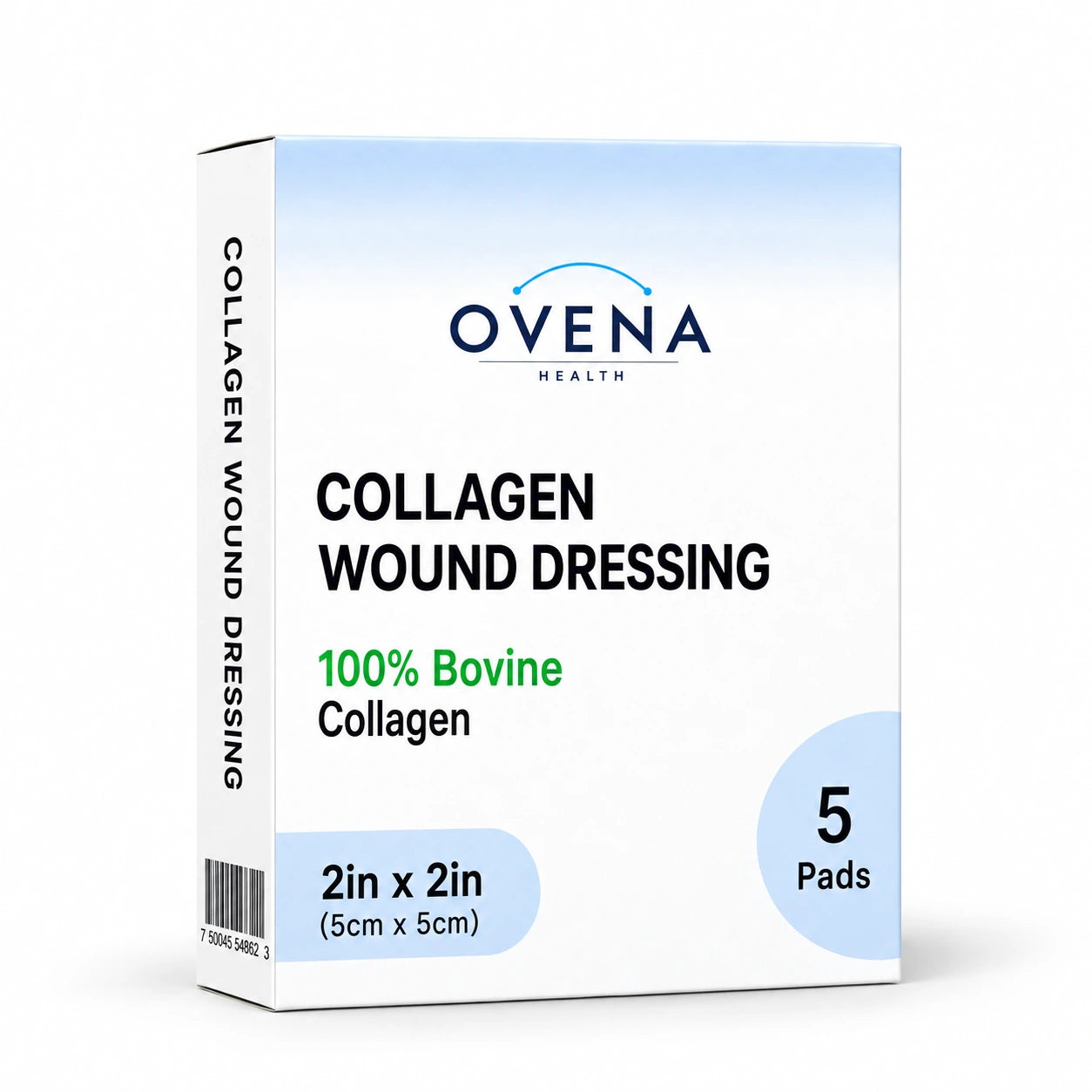
Collagen wound dressing for diabetic foot ulcer
Why diabetic foot ulcers stay stuck and how collagen addresses the specific biochemistry, plus the home care protocol, offloading, and glycemic control needed for healing.
 Reviewed byDavid Chahine, MD
In this guide
Why diabetic foot ulcers stay stuck
Diabetic foot ulcers (DFUs) precede 80% of lower-extremity amputations among people diagnosed with diabetes (1). Somewhere between 19% and 34% of people with diabetes are likely to develop a foot ulcer at some point, and roughly 40% of those who close one have another within a year (2). Healing DFUs is one of the highest-stakes problems in outpatient medicine, and the reasons they stay open are as much biological as procedural.
A DFU stays open for three reasons that compound:
- Persistent inflammation from hyperglycemia. High blood sugar drives chronic low-grade inflammation throughout the body. In the wound bed, this means inflammation never resolves — neutrophils and macrophages stay too long, producing damaging enzymes long past their useful purpose.
- Elevated matrix metalloproteinases (MMPs). MMPs are the enzymes responsible for tissue remodeling. In a normally healing wound they spike during inflammation and then drop. In a chronic wound they stay elevated — wound fluid taken from human pressure ulcers carries higher MMP levels and higher protease activity than fluid from acute surgical wounds (3). Excess protease activity degrades the new collagen the body is trying to lay down, so the wound never accumulates enough tissue to close. More on MMPs here.
- Compromised perfusion and neuropathy. Diabetes damages blood vessels and nerves. Less oxygen reaches the wound bed, less sensation means you keep walking on the ulcer without knowing it, and immune cells don't traffic in well. This one outranks the other two, and it gets its own section below.
Standard saline gauze does nothing to address any of these. It just covers the wound. That's why so many DFUs sit at the same size for months under "conservative" gauze care.
Circulation comes first
No dressing heals a wound that isn't getting blood. Peripheral arterial disease (PAD) sits behind roughly half of diabetic foot ulcers, and neuropathy hides the symptom that would normally announce it — cramping leg pain on walking. An ulcer on a foot with inadequate arterial supply will not close under collagen, under hydrocolloid, under foam, or under anything else, and no dressing choice compensates for the missing blood supply.
So before you settle into a home routine, have a clinician assess the circulation in that leg: pedal pulses, an ankle-brachial index (ABI), toe pressures, and a vascular referral if the study is abnormal or uninterpretable. Calcified vessels are common in diabetes and can push an ABI to a falsely reassuring number, which is one reason this belongs with a clinician rather than at home. If perfusion is the problem, restoring blood flow comes before dressing selection, not after it.
How collagen dressings address the DFU problem
A collagen wound dressing is a sterile sheet (or powder) of purified Type I bovine collagen, the same protein your body uses to build new skin scaffold. When you place it on a DFU, two things happen simultaneously:
- Sacrificial substrate. The MMPs in the wound bed degrade the dressing's collagen instead of the new tissue you are trying to grow. The dressing soaks up the enzyme load.
- Scaffold for granulation. The remaining collagen matrix gives fibroblasts, capillaries, and new tissue something physical to attach to and grow through.
The collagen is gradually absorbed as the wound heals. There's no peel-off — what is left of the old dressing comes away as a partial gel at the next change. Full application protocol here.
The protease-binding step has been shown outside the body. In an ex vivo study using wound fluid from people with diabetic foot ulcers, a collagen/oxidized regenerated cellulose (ORC) matrix bound and inactivated proteases, significantly reducing neutrophil elastase, plasmin, and MMP activity compared with wet gauze (4). Read that for what it is: a laboratory result rather than a healing outcome, run by the manufacturer of the product it tested, using collagen combined with ORC rather than a plain collagen sheet. It supports the mechanism. It does not tell you how fast your ulcer will close.
Clinical evidence
The most-cited trial is Veves et al. 2002, which randomized 276 people with diabetic foot ulcers to Promogran (a collagen/ORC dressing) or moistened gauze and followed them for up to 12 weeks. Complete closure came in at 37.0% with the collagen/ORC dressing vs 28.3% with gauze — a difference that did not reach statistical significance (P = .12), and the authors' own conclusion was that Promogran was comparable to moistened gauze. The one place a signal appeared was in ulcers open less than six months: 45% healed vs 33%, at P = .056, which the paper itself calls borderline (5).
Guidance is more cautious than most marketing copy suggests. The WOCN Society's clinical resource guide, listing options for wounds due to diabetes or neuropathic disease, says to "[c]onsider use of collagen or hyaluronic acid dressings that might promote healing" (6). Read the wording closely: consider, might, and collagen offered interchangeably with hyaluronic acid. That is an option a clinician may choose, not an instruction to use collagen. Other guideline bodies weigh the same evidence differently, so treat the choice as a conversation with your wound clinician rather than something to settle from an article.
What this means in plain language: a collagen dressing is not a shortcut. Roughly 30-40% of diabetic foot ulcers are healed at 12 weeks (1), and the ones that get there do so on offloading, circulation, infection control, and glucose management at least as much as on any dressing. Collagen addresses one specific problem — the protease load in the wound bed. It is worth using for that reason, with your clinician's agreement, and not as a substitute for the rest of the plan.
The home care protocol
The standard protocol your wound care nurse will hand you. Adapt to whatever your specific clinician prescribes.
- Wash your hands with soap and water. Put on clean nitrile gloves.
- Inspect the foot end-to-end. Look at the wound, the surrounding skin, the entire foot top-and-bottom, between the toes, and the heel. Neuropathy hides new injuries — that is exactly why daily inspection is non-negotiable.
- Irrigate the wound with sterile saline. Squeeze the bottle to flush — don't just dab. Do not use hydrogen peroxide or alcohol. Iodine does not belong on granulating tissue either, though a clinician may deliberately use it to keep dry, stable eschar dry — that is their call to make, not a home decision.
- Pat the surrounding skin dry. The wound bed stays slightly moist.
- Apply the collagen sheet trimmed to the wound size, with about a quarter inch of overlap. If the ulcer tunnels or undermines — a probe slides into a tract or under the skin edge — stop and be seen the same day. A tract is not something to pack at home, and powder is not the answer to it.
- Cover with a non-adherent secondary dressing. A foam works well for DFUs with moderate drainage. Secure it with hypoallergenic tape or a wrap.
- Put the offloading device back on (cast walker, postoperative shoe, total contact cast). Walking on the foot without offloading undoes everything you just did.
- Change every 2-3 days, or on whatever schedule your clinician set. Closer to every day for high drainage, closer to every 3 for quiet, low-drainage wounds.
- Never soak the foot. Soaking does not clean a wound. It macerates the skin around it, and in a neuropathic foot a soak is a common route to a burn or a fresh ulcer.
- Never use a heating pad, hot water bottle, or electric blanket on the feet. Neuropathy means you will not feel a burn until after it has happened.
The other half: offloading and glucose control
You can use the best dressing in the world and the wound won't heal if you keep walking on it. Offloading is the single most important factor in DFU healing after addressing infection. The standard hierarchy:
- Total contact cast (TCC) — the most effective offloading, and the reference standard. Requires regular replacement by a clinician.
- Removable cast walker (a CAM boot, for example) — nearly as effective as a TCC when it is worn consistently, and less effective in practice precisely because it comes off.
- Felted-foam pads plus a postoperative shoe — entry-level offloading, useful when more rigid options aren't tolerated.
Equally critical: blood glucose control. It belongs in the healing plan alongside the dressing, and most wound centers coordinate with your endocrinologist or primary care clinician to work on it in parallel with wound care.
Warning signs that need emergency care
- Fever, chills, or feeling generally unwell with an open foot wound
- Redness spreading outward from the wound, or skin that is hot and tight
- You can see bone, tendon, or fat in the wound, or a probe can touch bone
- Foul-smelling discharge (yellow, green, or gray with odor)
- New black tissue at the wound edges or center
- Sudden increase in pain, or new pain in a foot that normally feels nothing
- Drainage that is climbing from one change to the next instead of settling
Diabetic foot infections can become limb-threatening within 24-48 hours. An emergency department can image the foot, draw labs, and start intravenous antibiotics in one visit; urgent care generally cannot do any of the three. Call your podiatrist or wound clinician on the way if you can, but don't let a callback decide whether you go.
Call your wound clinician within a day or two — not an emergency, but not "wait for the next appointment" either — if the ulcer has not changed at all after two weeks of consistent care, or if it has now been open longer than four weeks. A wound open more than four weeks needs an assessment rather than another month of the same dressing: circulation, infection, offloading, and a biopsy if your clinician thinks one is indicated. Collagen can be part of the plan your clinician sets from there.
Realistic healing timelines
What to expect with consistent care, adequate circulation, offloading, glucose control, and collagen dressings:
- Week 1-2: Slough lifts from the wound bed and new pink or red granulation tissue appears. Drainage should hold steady or fall. Rising drainage is a sign of infection, not a sign that a wound is "waking up" — if the secondary dressing needs changing more often this week than last, call your clinician.
- Week 3-6: Granulation fills the wound depth. The wound starts contracting from the edges inward. Visible wound area starts shrinking.
- Week 6-12: Epithelialization — new skin migrating across the wound surface from the edges. The wound continues to shrink. Ulcers that close with conservative care usually do so in this window; across diabetic foot ulcers overall, roughly 30-40% are healed at 12 weeks (1).
- Beyond 12 weeks: If the wound hasn't closed by 12 weeks despite consistent care, escalate. That means a wound center referral for advanced therapy: negative pressure wound therapy, hyperbaric oxygen, skin substitutes, or biologic dressings.
Photograph the wound at every dressing change. Objective comparison beats memory, and week-over-week progress is often easier to see in two photos than in the wound in front of you.
Ovena collagen for DFU care
Our collagen dressings and powder are FDA 510(k)-cleared, the same regulatory class wound clinics use. Itemized receipts with the HCPCS code on request (A6021 for our 2x2 and 4x4 sheets, A6010 for the powder). These items are commonly FSA/HSA eligible; your plan administrator decides, and a letter of medical necessity may be required. Free shipping over $75.
Shop collagen wound dressings → Shop the complete kit →Frequently asked questions
How long does it take a DFU to heal with collagen?
Should I use collagen on an infected DFU?
Can I use collagen dressings at home for my DFU?
Is offloading really that important?
What HCPCS codes apply to collagen dressings?
Sources
- Armstrong DG, Tan TW, Boulton AJM, Bus SA. Diabetic foot ulcers: a review. JAMA. 2023;330(1):62-75.
- Armstrong DG, Boulton AJM, Bus SA. Diabetic foot ulcers and their recurrence. N Engl J Med. 2017;376(24):2367-2375.
- Yager DR, Zhang LY, Liang HX, Diegelmann RF, Cohen IK. Wound fluids from human pressure ulcers contain elevated matrix metalloproteinase levels and activity compared to surgical wound fluids. J Invest Dermatol. 1996;107(5):743-748.
- Cullen B, Smith R, McCulloch E, Silcock D, Morrison L. Mechanism of action of PROMOGRAN, a protease modulating matrix, for the treatment of diabetic foot ulcers. Wound Repair Regen. 2002;10(1):16-25. Ex vivo study; the authors were employed by the manufacturer of the dressing tested.
- Veves A, Sheehan P, Pham HT. A randomized, controlled trial of Promogran (a collagen/oxidized regenerated cellulose dressing) vs standard treatment in the management of diabetic foot ulcers. Arch Surg. 2002;137(7):822-827.
- WOCN Society. Lower-Extremity Wounds Due to Venous Disease, Arterial Disease, or Diabetes Mellitus and/or Neuropathic Disease: Clinical Resource Guide. Revised December 2021.